![]()
![]()
![]()
![]()
![]()
Colour Coding:
Image indicators are shown onscreen, as coloured bars above and below the teeth, to show there are images present.
Image Age:
To differentiate images on the basis of age, the older they get, the paler the coloured indicator bars will become, allowing the dentist to see at a glance how old (and therefore how relevant) an image is.
![]()
![]()
![]()
![]()
![]()
Indicator bar location for Bitewing and Panoral
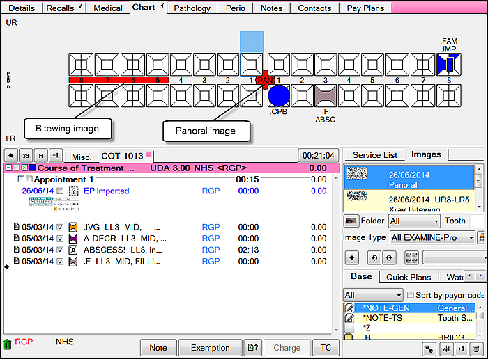
Where the tooth range covers both jaws (a bitewing) it is represented as a single bar between the rows of teeth spanned.
A panoral image is located centrally.
Other Details:
Where there are multiple images for a tooth, the bars will be drawn from oldest to newest, so that the newest, most relevant images are shown on top.
Indicator bars are drawn so that they span the teeth that the image belongs to; e.g. an image covering teeth LL5 to LL8 will be drawn as a single rectangle alongside LL5 through LL8 on the chart.
An image indicator is displayed only if the teeth it covers are on the chart; e.g. deciduous tooth x-rays are not drawn unless the the deciduous teeth are being displayed. Where mixed dentition is being displayed, then the image indicators for the deciduous and permanent teeth are shown in the same place, as this is most likely to be the case, as in a retained deciduous tooth alongside the permanent tooth. For example, an indicator bar for an x-ray for UL5 would be drawn at the same location as one for ULE.
Example:
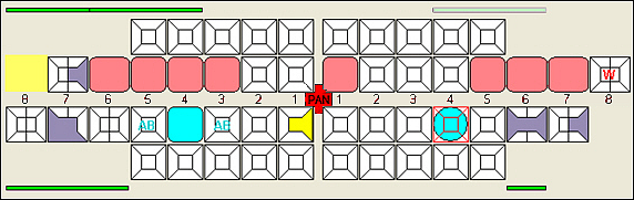
In the example above, there are the following images associated with the chart: